Exams
Full Ocular Health ExamsOur professional eye doctors will perform an extensive eye examination which includes vision testing, and a thorough assessment of your eye health and coordination.
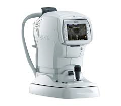
Vision testing of your visual status with your present glasses, your contact lenses or simply your own eyes is the first important step in your examination. Then using advanced equipment, we will do a series of lens tests (called a refraction) to determine if you need glasses or contact lenses to improve your vision. By helping you see your best, in many cases we can enhance your academic,occupational or recreational performance. All complete examinations include an eye health assessment with various instruments including an ophthalmoscope and biomicroscope. These instruments allow the doctor to examine the internal structures of the eye, including the optic nerve, retinal blood vessels and the retina in general. In addition to checking for cataracts and glaucoma, many systemic diseases such as high blood pressure and diabetes have signs that can be detected by a thorough examination of the eyes. We also perform a painless procedure called tonometry to measure the pressure inside your eye or intraocular pressure (IOP). This common test is important in the detection of glaucoma. |
In case of an emergency please call the office, as we will try our best to accommodate an appointment for you immediately. If the office is closed please proceed to the hospital emergency or walk-in clinic. Thank you.
Supplementary Testing
The optometrist may require that you have supplementary diagnostic testing in order to assist the doctor in monitoring and managing current and potential eye health risks or complications. Some testing may come at an additional cost and others may be fully covered by OHIP, please ask the doctor or staff member if you think you may require any additional testing and whether it is covered by OHIP.
|
Optical Coherence Tomography (OCT)
An OCT is a non-invasive imaging method, which is essentially an ultrasound of your eye, but instead of sound waves the OCT uses the reflection of light. It is through the use of OCT that optometrists routinely diagnose macular diseases such as neovascularization, diabetes, and age-related macular degeneration.
|
Retinal Photographic Documentation (RPD)
Retinal imaging takes a digital picture of the back of your eye. It shows the retina, the optic disk, and blood vessels. This helps your optometrist check the health of your eyes and screens for the potential onset of complications; such as, glaucoma, macular degeneration, high cholesterol, and diabetic retinopathy.
|
Pachymetry
A pachymeter is a medical device used to measure the thickness of the eye's cornea. Corneal pachymetry is useful in determining the risk of developing glaucoma and interpreting unexpected intra-ocular pressure (IOP) measurement results.
|
Automated Visual Fields Analysis
The test involves a presentation of lights to an individual eye. The minimum light level detected by the patient for each presented point is used to assess the health of the visual pathway, from the eye to the back of the head.
Visual field analysis is one of the tests required for the diagnosis and ongoing treatment management of glaucoma. It is also useful in the diagnosis of neurological disorders. Patients at high risk of glaucoma, having neurological signs, headaches, or unusual symptoms will benefit from such testing. |


